
Ai in Custom Facial Implant Surgery
How Useful is AI-Generated Imaging in Custom Facial Implant Designs?
Artificial intelligence-generated imaging is beginning to influence nearly every aspect of aesthetic medicine, and custom facial implant design is no exception. While AI imaging tools have become highly visible through consumer applications that can instantly simulate facial changes, their true usefulness in custom facial implant surgery is more nuanced. AI-generated imaging can be extremely valuable as a communication, visualization, and planning tool, but it also has important limitations that patients must understand.
The Evolution of Facial Implant Design
Traditional facial implant surgery has historically relied on stock implants with intraoperative modification. While effective for many patients, stock implants have limitations because human facial anatomy varies considerably. Over the last decade, the rise of 3D CT imaging and CAD/CAM (computer-aided design/computer-aided manufacturing) technology has transformed facial implant surgery by allowing implants to be designed specifically for an individual patient’s skeletal anatomy.
Custom facial implants are now commonly designed using:
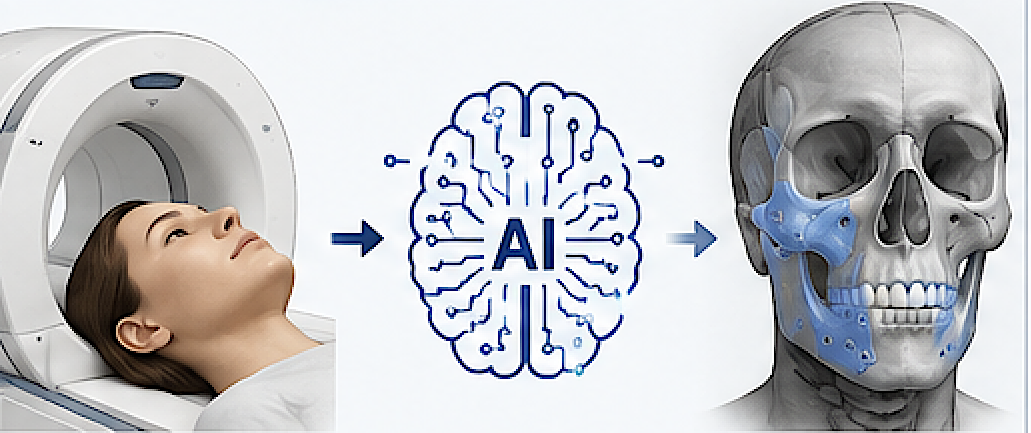
- High-resolution CT scans
- 3D skeletal reconstructions
- Virtual surgical planning software
- CAD-based implant engineering
- 3D printing and milling technologies
The addition of AI-generated imaging represents the next stage in this evolution.
What is AI-Generated Imaging?
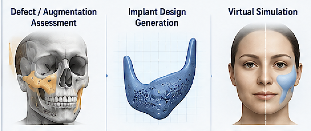
AI-generated imaging refers to software systems that use machine learning models to create, predict, simulate, or modify images based on large datasets and pattern recognition. In facial implant surgery, AI imaging may be used in several different ways:
- Predictive facial outcome simulation
- Automated facial analysis
- Soft tissue response prediction
- Morphologic enhancement suggestions
- Automated implant contour generation
- Symmetry correction analysis
- Virtual patient communication tools
These applications range from highly useful to currently experimental.
The Most Valuable Use: Patient Communication
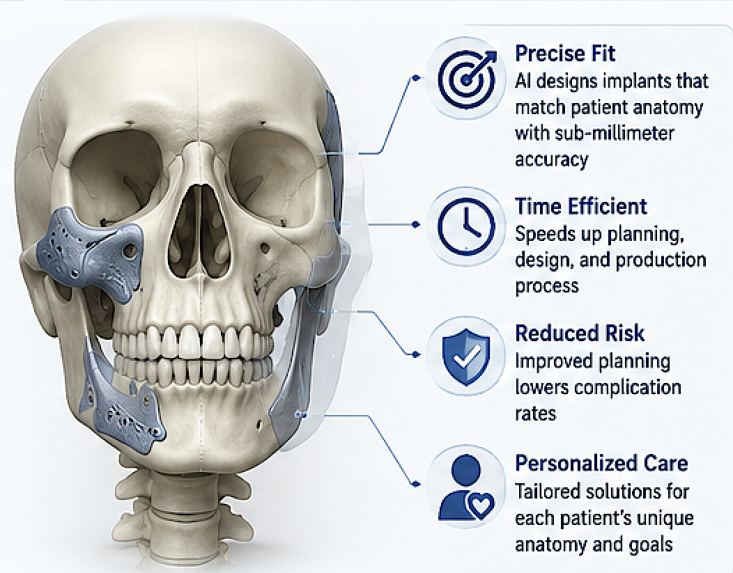
While there are some distinct benefits for Ai in custom facial implants, its greatest current usefulness of AI is probably patient communication and expectation alignment.
Many patients struggle to visualize skeletal changes from implant surgery. Traditional before-and-after photos may not accurately represent their anatomy or desired outcome. AI imaging allows surgeons to generate approximations of possible aesthetic changes in a more personalized way.
For example, AI-assisted simulations can help demonstrate:
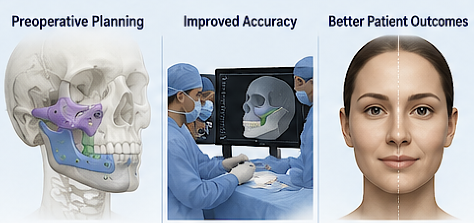
- Chin projection changes
- Jawline widening
- Midface augmentation
- Infraorbital contour improvements
- Facial balance adjustments
- Symmetry corrections
This can improve the consultation process by helping patients articulate goals more clearly.
However, these simulations should never be interpreted as exact predictions.
AI is Better at Surface Visualization Than Structural Design
One important distinction is that AI-generated facial images are generally better at simulating surface appearance than actually engineering implants.
Custom facial implants require:
- Precise skeletal fit
- Bone interface conformity
- Attention to nerve pathways
- Soft tissue thickness considerations
- Surgical access limitations
- Fixation planning
- Long-term biomechanical stability
These are engineering and surgical problems rather than purely visual problems.
While AI can generate aesthetically appealing images, implant design still depends heavily on surgeon judgment, anatomical expertise, and biomedical engineering.
Currently, AI is far more useful as a visualization assistant than as a replacement for custom implant design expertise.
Soft Tissue Prediction Remains the Biggest Challenge
One of the most difficult aspects of facial implant surgery is predicting how soft tissues will respond to skeletal augmentation.
Bone movement and implant dimensions are measurable. Soft tissue response is much harder to predict because it varies according to:
- Skin thickness
- Age
- Fat distribution
- Muscle tone
- Tissue elasticity
- Scar history
- Previous surgery
- Ethnicity
- Weight fluctuations
AI systems are improving in soft tissue prediction by learning from large surgical datasets, but the variability between patients remains substantial.
This is particularly true in:
- Midface augmentation
- Paranasal implants
- Infraorbital implants
- Combined jawline procedures
- Revision surgery
As a result, AI-generated imaging should still be viewed as illustrative rather than predictive.
AI and Symmetry Analysis
One area where AI is becoming genuinely powerful is facial asymmetry analysis.
Facial asymmetry is common and often difficult to analyze manually. AI systems can:
- Detect skeletal asymmetries
- Quantify contour discrepancies
- Compare bilateral dimensions
- Identify occlusal canting
- Map volumetric deficiencies
- Generate mirrored reference anatomy
This can significantly improve custom implant planning, especially in:
- Congenital asymmetry
- Hemifacial microsomia
- Post-traumatic deformities
- Revision facial implant surgery
- Orthognathic surgery adjuncts
In these cases, AI-assisted imaging may improve surgical precision and implant customization.
AI-Assisted Implant Design
Some emerging software platforms now incorporate AI-assisted implant generation.
These systems may suggest implant contours based on:
- Idealized facial proportions
- Population datasets
- Mirror-image anatomy
- Surgeon design preferences
- Historical case libraries
Potential benefits include:
- Faster design workflows
- More efficient contour generation
- Improved consistency
- Automated smoothing and transitions
- Better adaptation to complex anatomy
But surgeon oversight remains critical.
An aesthetically pleasing implant on a computer screen may still be surgically impractical or anatomically unsafe.
Limitations of AI Imaging in Facial Implants
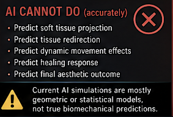
Despite its promise, AI-generated imaging has significant limitations.
1. Overpromising Results
Consumer AI applications can create unrealistic expectations. Patients may believe AI-generated outcomes are guaranteed surgical results when they are merely approximations.
2. Lack of Biological Understanding
AI systems may not fully account for:
- Tissue healing
- Scar contraction
- Implant settling
- Edema
- Muscle movement
- Aging changes
- Long-term adaptation
3. Dataset Bias
AI models are only as good as the datasets they are trained on. Limited diversity in age, ethnicity, gender, or facial structures can reduce accuracy.
4. Surgical Constraints
AI-generated idealized outcomes may not consider:
- Surgical exposure limitations
- Nerve safety
- Existing scar tissue
- Bone thickness
- Occlusal issues
- Implant fixation constraints
5. Revision Complexity
Revision surgery remains especially difficult for AI systems because previous surgery alters anatomy unpredictably.
Where AI Will Likely Become Most Important
The future usefulness of AI in custom facial implants will likely expand in several areas.
Automated Anatomical Mapping
AI will increasingly automate segmentation of:
- Bone
- Soft tissue
- Nerves
- Sinuses
- Dental roots
- Vascular structures
This could greatly reduce planning time.
Predictive Soft Tissue Modeling
As datasets improve, AI may become substantially better at predicting how skin and soft tissues respond to skeletal augmentation.
Real-Time Implant Iteration
Future systems may allow surgeons and patients to interactively adjust implant dimensions with immediate visualization of predicted outcomes.
Outcome-Based Learning
AI systems may eventually learn from thousands of postoperative scans and outcomes to refine implant recommendations.
The Surgeon Still Matters Most
Despite advances in AI imaging, successful custom facial implant surgery still depends primarily on:
- Surgical judgment
- Aesthetic vision
- Anatomical expertise
- Experience with soft tissue behavior
- Intraoperative decision-making
- Understanding patient psychology
AI can assist the process, but it cannot replace the surgeon’s ability to interpret anatomy, manage complications, or create individualized aesthetic outcomes.
The most successful future model will likely be a hybrid approach where:
- AI enhances visualization and analysis
- CAD technology enables precise implant fabrication
- Surgeons provide final design judgment and execution
Conclusion
AI-generated imaging is already useful in custom facial implant design, particularly for patient communication, asymmetry analysis, visualization, and surgical planning support. Its greatest strength currently lies in helping patients and surgeons better understand potential aesthetic changes.
However, AI remains far less reliable in predicting exact surgical outcomes or independently designing implants without expert oversight. Facial implant surgery involves complex interactions between bone, soft tissue, surgical technique, healing biology, and aesthetics that AI cannot yet fully replicate.
As machine learning systems continue to evolve and incorporate larger surgical datasets, AI will likely become an increasingly valuable adjunct in custom facial implant surgery. But at present, it should be viewed as a sophisticated support tool rather than a replacement for experienced surgical planning and judgment.
Ultimately, the best results still come from combining advanced imaging technology with experienced surgeon-driven design principles.
The Current Limitations of Ai in Custom Facial Implant Surgery
AI still cannot accurately predict soft tissue changes in custom facial implant surgery because the problem is fundamentally a highly nonlinear biologic mechanics problem with incomplete data and too many patient-specific variables. The implant itself is geometrically predictable; the soft tissue response is not.
The limitations fall into several categories:
1. Soft tissue is not a uniform material
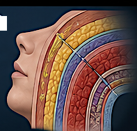
Bone is rigid and mathematically stable. Facial soft tissue is not.
The face contains:
- skin
- fat compartments
- fascia
- muscle
- ligaments
- periosteum
- scar tissue
- varying hydration and elasticity
Each layer behaves differently under implant-induced stretch and projection. Two patients receiving the exact same implant can show noticeably different external outcomes.
AI models work best when:
- the system is deterministic
- the variables are measurable
- the physics are consistent
Facial soft tissue is none of those.
2. The relationship between implant shape and visible change is nonlinear
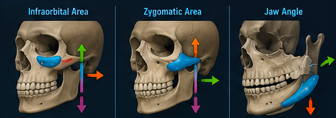
A common misconception is:
“3 mm of implant projection = 3 mm of visible soft tissue projection.”
In reality:
- some areas translate nearly 1:1
- others dampen the effect
- others redirect tissue vectors entirely
For example:
- infraorbital implants may create anterior projection, lower eyelid support, or lateral widening depending on tissue tethering
- jaw implants may increase width more than projection in one patient, but mostly vertical definition in another
Small geometric changes can produce disproportionately different aesthetic outcomes.
3. Facial retaining ligaments strongly affect tissue movement
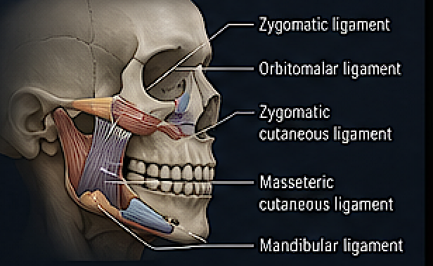
The face is compartmentalized by retaining ligaments:
- zygomatic ligaments
- mandibular ligaments
- orbitomalar ligaments
- masseteric cutaneous ligaments
These create “fixed points” that resist predictable deformation.
AI currently has poor ability to infer:
- ligament stiffness
- tethering strength
- tissue release characteristics
from standard imaging.
CT scans show bone well, but not dynamic soft tissue biomechanics.
4. Dynamic muscle movement changes the result
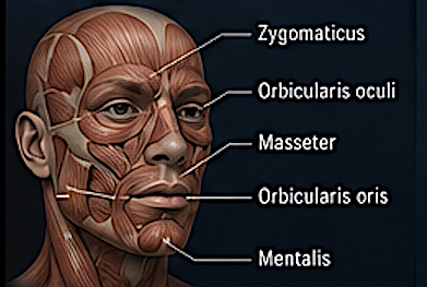
The face is not static.
Soft tissue appearance changes during:
- smiling
- speaking
- chewing
- animation
- head positioning
An implant interacts with muscles like:
- mentalis
- masseter
- zygomaticus
- orbicularis oris
AI prediction systems usually rely on static scans, not real-time biomechanical animation models.
A realistic predictive model would need:
- high-resolution anatomy
- dynamic muscle simulation
- individualized tissue elasticity
- neuromuscular behavior
That is computationally and biologically difficult.
5. Healing response is biologically unpredictable
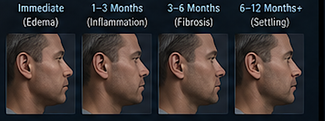
Postoperative changes include:
- edema
- fibrosis
- capsule formation
- tissue relaxation
- scar contracture
- implant settling
These vary widely between patients.
AI struggles because healing is influenced by:
- genetics
- age
- vascularity
- smoking
- prior surgery
- inflammatory response
- hormonal state
These variables are not adequately captured in current datasets.
6. There is not enough high-quality training data
AI prediction depends on massive standardized datasets.
Facial implant surgery lacks:
- standardized implant designs
- standardized photography
- standardized lighting
- consistent head positioning
- long-term follow-up scans
- paired CT + soft tissue outcome datasets
Most surgeons also use highly individualized designs, making generalized learning difficult.
Compared with fields like autonomous driving or dermatology imaging, the dataset size is tiny.
7. Aesthetic outcomes are partly subjective
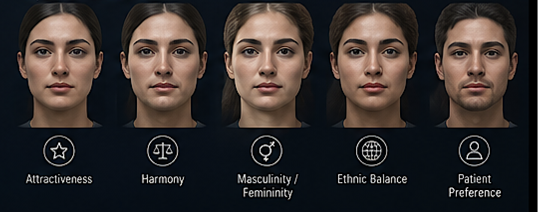
Even if AI perfectly predicted tissue movement, it still would not fully solve:
- attractiveness
- harmony
- masculinity/femininity perception
- age perception
- ethnic facial balance
- patient satisfaction
These are perceptual and cultural variables rather than purely geometric ones.
8. Current AI models are mostly geometric, not biomechanical
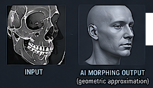
Most current “AI facial simulation” systems are:
- morphing systems
- statistical averaging systems
- image warping tools
They are not true finite-element biomechanical simulations.
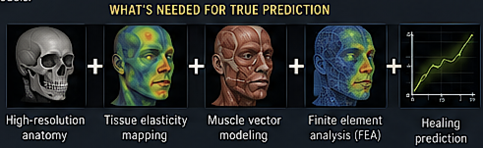
A true predictive engine would require combining:
- finite element analysis (FEA)
- tissue elasticity mapping
- muscle vector modeling
- biologic healing prediction
- machine learning
That integration is still immature.
What AI can do reasonably well today
AI is becoming useful for:
- implant sizing suggestions
- symmetry analysis
- cephalometric analysis
- identifying deficient skeletal zones
- generating probable aesthetic ranges
- surgical planning assistance
- implant design optimization
But not precise soft tissue forecasting.
What will likely improve prediction in the future
The biggest advances will probably come from combining:
- High-resolution 3D facial scans
- Dynamic motion capture
- MRI-based soft tissue characterization
- Patient-specific finite element models
- Large longitudinal surgical datasets
- Physics-informed neural networks
Eventually AI may provide:
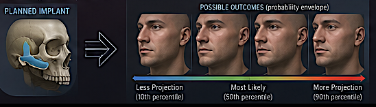
“probabilistic soft tissue outcome envelopes”
rather than exact predictions.
That is likely more realistic than expecting a single perfectly accurate postoperative face simulation.